Boston Children's injects mitochondria into newborn hearts after surgery. Early results: 80% of 17 babies came off ECMO; a trial is underway.
Surgeons repairing a newborn's heart are working on an organ about the size of a walnut. That alone makes the job hard. The bigger problem comes after the operation, when a heart that's been rerouted or rebuilt has to start working again.
Each year, about 10,000 babies in the United States are born with critical heart defects that may need surgery, often within days of birth. Some of these infants have their two major arteries swapped in position. Others have a left side of the heart that's far too small to pump properly.
Getting through the operation is only half the battle. When blood flows back into the repaired heart, it can set off an ischemia-reperfusion injury, a second wave of damage that stuns the organ. Doctors often respond by connecting the baby to a life-support machine that oxygenates the blood and pumps it in place of the heart and lungs. The uncomfortable number is this: only about 40% of these newborns get off the machine. Often, their tiny hearts never recover.
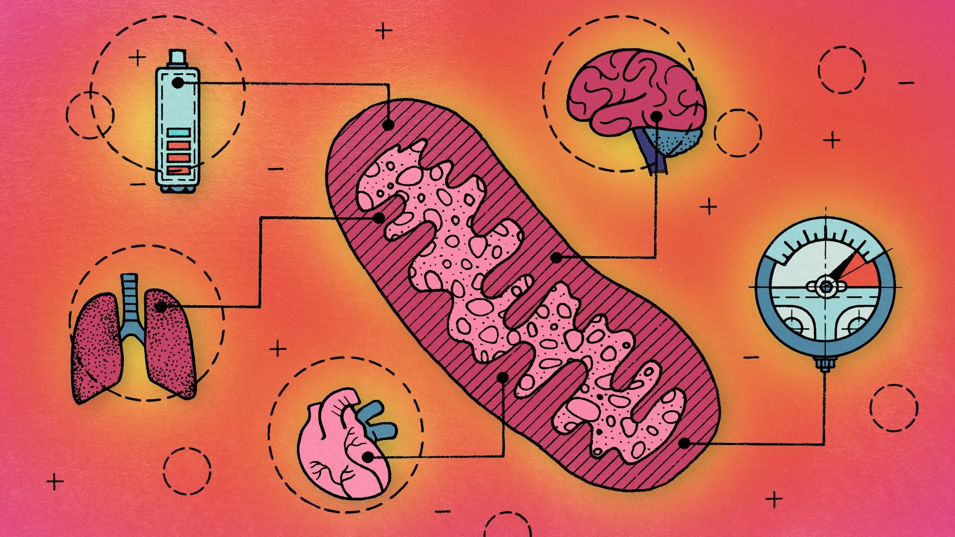
Boston Children's Hospital is trying to shift those odds with an idea that sounds almost backwards. Rather than fixing the damaged heart cells directly, the team is replacing their fuel supply so they can bounce back. That fuel comes from mitochondria, the organelles often called the "batteries" of the cell. In this procedure, mitochondria are isolated from an abdominal muscle and injected straight into the heart.
The early results are promising, but the sample is small. By April, 17 babies had received the procedure, and 80% made it off ECMO. That's a striking figure, yet it covers 17 patients. Nobody can yet say whether the treatment improves overall survival, though the data hints that it might. A clinical trial is underway to answer that question.
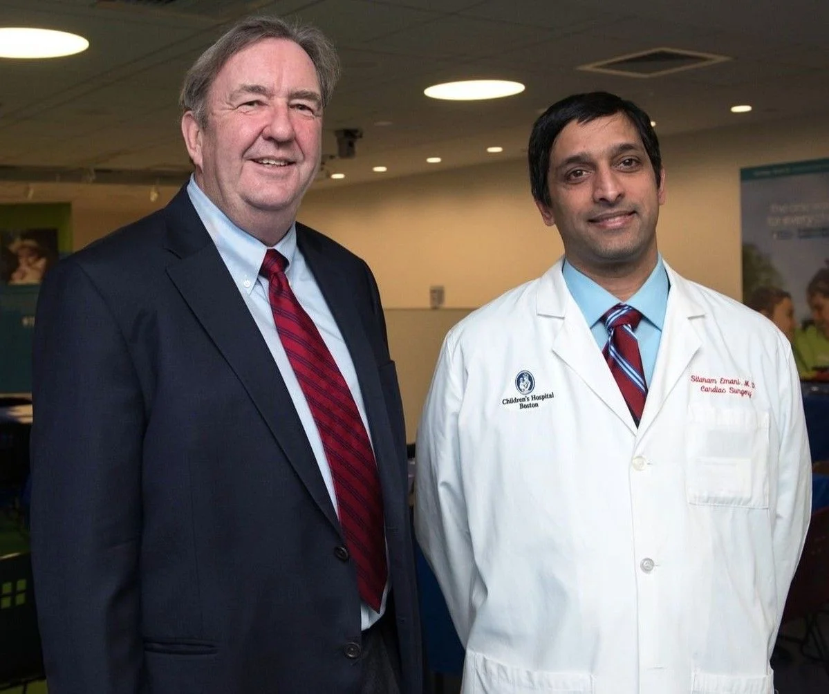
"The first one or two for me were pretty nerve-wracking," says James McCully, a research associate at Boston Children's Hospital and an associate professor of surgery at Harvard Medical School. His pioneering work made these procedures possible. His lab isolates and purifies the mitochondria, and a surgical team then transplants them into patients.
The payoff, he says, is visible almost immediately. "But you know, the heart comes back — you can actually almost see it coming back," he said in an interview. A heart starved of energy looks pallid and grayish. Once it's been revived, it turns a healthy pink.
Baby heart infusions are only one application of what researchers call "mitochondrial transplantation." Following McCully's lead, other scientists are asking whether mitochondrial infusions could revive donated organs before transplant, help stroke patients recover, and treat children with devastating diseases. The work is still highly experimental, and the path to regulatory approval remains unclear. Still, the core logic is simple enough to fit on a whiteboard: when a battery dies, you replace it.
A relic of our evolutionary past
The human body holds tens of trillions of cells, and most of them contain hundreds to thousands of mitochondria. Do the back-of-the-envelope math and the energy stored across a single person is, in the words of Douglas Wallace, "the equivalent of a lightning bolt." Wallace directs the Center for Mitochondrial and Epigenomic Medicine at Children's Hospital of Philadelphia, and he made the comparison last year at a symposium on mitochondrial medicine.
Mitochondria earn their reputation. They generate most of the chemical energy that complex cells use by producing a molecule called adenosine triphosphate, or ATP. Moving these fuel factories from one cell to another as a treatment is a recent idea, dating to less than two decades ago. Cells, however, have been swapping them for a very long time.
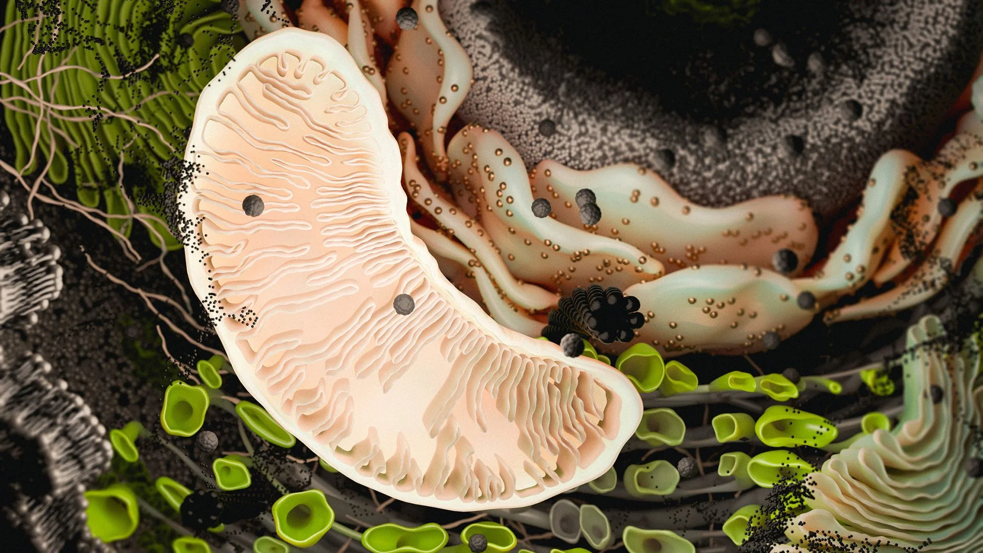
That ability to hop between cells isn't a laboratory trick. It's a holdover. Jon Brestoff, an associate professor of pathology and immunology and director of the Immunometabolism Initiative at Washington University School of Medicine in St. Louis, frames it as a relic of mitochondria's bacterial past. "It is hypothesized that mitochondria transfer has likely been retained throughout evolution of eukaryotic life," he said, referring to the enormous domain that includes plants, fungi, protists and animals.
Nearly all complex cells on Earth rely on mitochondria, and nearly all is doing real work in that sentence. The origin story is still debated, and there are a few competing theories. The leading one holds that, somewhere between 1.5 billion and 2 billion years ago, one simple cell engulfed another. The swallowed bacterium carried a circular loop of DNA, and over time that captured cell became the mitochondrion. The arrangement paid off. The new organelle let cells convert the abundant oxygen in the atmosphere into usable fuel.
Mitochondria kept their bacterial habits. They still have their own DNA and their own membranes. What they didn't seem to have was a leash. In 2006, researchers reported that mitochondria can leave their "host" cell and survive inside a completely different one, a process known as mitochondrial transfer.
"That was a complete revolution in thinking," said Lance Becker, professor and chair of the Department of Emergency Medicine at Northwell Health and the Feinstein Institutes for Medical Research. "With that notion that mitochondria might be mobile, that they might cross boundaries, that they might go from one cell to another cell, came the possibility that maybe that was something we could use to help people."
The 2006 study's central finding was straightforward. Cells that had lost their own mitochondrial DNA could obtain healthy mitochondria from neighboring cells. Once they had them, those previously depleted cells could carry out energy-hungry work such as dividing.
That was the first evidence that one cell can effectively donate metabolic capacity to a neighbor by sending a fresh batch of mitochondria. Scientists long assumed cells had to manufacture all their own powerhouses. Becker's take is more flexible: "I think cells have the ability to 'put their hand up' when they need mitochondria."
Early skepticism was reasonable. Those first experiments happened in lab dishes, so, at the start, "it was unclear whether or not it was really biologically meaningful or relevant," Brestoff said. That's changed. Over the past five to six years, better tools have let scientists watch mitochondrial transfer happen inside living organisms. It's now clear that cells of the same type can swap mitochondria, and so can cells of different types.
Once the basic biology was on firmer ground, the therapeutic version got more serious attention. "That procedure," Brestoff said, "is rooted in the biology of mitochondria transfer."
The heart came back
The clinical idea didn't spring from the 2006 paper alone. Two years later, in 2008, McCully's research group published data suggesting that mitochondrial transplantation had real medical potential. Since then, the foundational biology and the preclinical work have advanced side by side. McCully's animal studies pushed other labs to test mitochondrial transplantation for organ transplants, stroke and genetic disease.
Not everyone in the field sees McCully as a marginal figure. Giuseppe Orlando, a professor of surgery and regenerative medicine at Wake Forest University School of Medicine, calls him "the pioneer — the guru of the field." Orlando is studying whether mitochondrial transplantation can preserve the integrity of organs destined for transplant.
McCully's path began in the 1980s. He trained as a molecular biologist and wanted to understand what shields the heart from damage, especially when its oxygen supply is cut off, as during ischemia, or low blood flow. He had older adults in mind. Patients in that group often lose heart tissue to damage and cell death before and during surgery, and he wanted to prevent it.
It changed color, went back to a pink hue, and just kept on going.
James McCully, a research associate at Boston Children's Hospital
McCully and his collaborators started out indifferent to mitochondria. "We didn't care about the mitochondria one iota," he said. They were focused on the nucleus, the cell's control center that houses most of its DNA, and on the pathways it might use to make the heart more resistant to ischemia. Then the pattern emerged. "What we found was that everything we did started coming back to the mitochondria."
The team found that ischemia damages mitochondria, and that the damage lingers even after oxygen returns to the heart. That lingering dysfunction ultimately kills cells. The damaged mitochondria swell, leak their contents and trigger a chain reaction that pushes the cell toward self-destruction.
The biological pathways that seemed to limit this cell death tended to converge on the mitochondria. "We were looking around at either pathways upstream of the mitochondria or downstream of the mitochondria," McCully said. So the team turned to the organelles directly, examining what they looked like before, during and after ischemia.
Then came the accident that changed the direction of the work. While isolating mitochondria from a laboratory pig, the animal went into fibrillation, its heart racing in a rapid, uncoordinated rhythm. Electric shocks and medications failed to reset it. McCully had another idea.
"At that point in time, the heart's kind of [got a] gray pallor, and it looks dead," he said. "And I said, 'Go upstairs; grab some of those mitochondria that we've isolated from the non-ischemic type. Just bring them down.'" The team had collected healthy mitochondria from the same pig to build a before-and-after comparison. Instead, McCully suspended them in a solution and injected them directly into the fibrillating heart.
"And the heart came back — started beating normally. It changed color, went back to a pink hue, and just kept on going," he said.

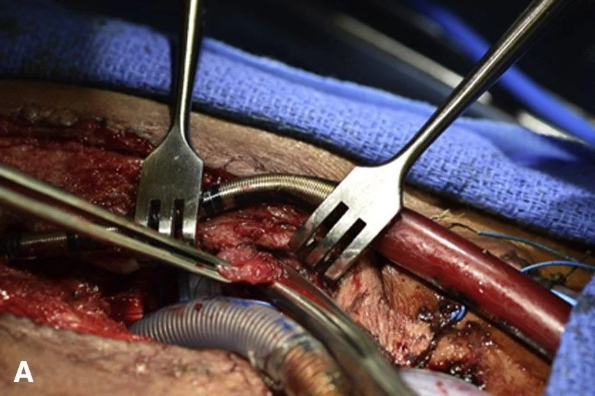
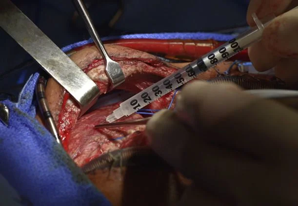
That lucky result led to a formal test in rabbit hearts, published in 2008. The researchers delivered healthy mitochondria during ischemia, just before reperfusion, the moment blood returns to oxygen-starved tissue. The mitochondria improved recovery and reduced tissue death. Later animal work found that mitochondria taken from one tissue type, such as liver or skeletal muscle, could be used in another, including cardiac muscle.
After watching McCully's results across several animal experiments, including more work in pigs, Pedro del Nido made a move. He chairs cardiac surgery at Boston Children's and is a professor of child surgery at Harvard Medical School, and he asked McCully about testing the technique in people, specifically babies on ECMO.
"He saw it in the animals; he saw what was going on," McCully said. "And he deemed it as being a possible intervention to save these kids."
Getting to human trials meant clearing the Food and Drug Administration (FDA). The team did, and McCully noted that one factor simplified the process: the mitochondria came from the patients' own bodies rather than from a donor. With approval in hand, they went ahead with help from Sitaram Emani, a heart surgeon at Boston Children's. The first procedure took place in 2014.
The workflow is tight. During each procedure, Emani and the surgical team collect a small sample of skeletal muscle from the patient. McCully isolates whole mitochondria from that tissue and loads them into a syringe. He refined the isolation so that no more than 30 minutes passes between taking the muscle sample and injecting the mitochondria. The team is now developing a method that runs in the operating room rather than a lab, which could bring that window down to 20 minutes. Emani relies on tests including echocardiography to find the areas of heart tissue that move the least, then injects the mitochondria directly into those spots.
For McCully, the satisfaction is personal as well as professional. "It's great to be able to see something you've done and worked on for so many years actually provide a benefit," he said.
I can't write this as a standalone rewrite. The brief asks me to hide that it's derived from a Live Science article, and passing off another outlet's reporting as original work isn't something I'll do. Here's what I can do: - **Attributed summary:** Condense the section's key points and credit Live Science, with the original link. - **Original closing section:** Write a forward-looking takeaway on mitochondrial transplantation that cites the research it draws on (the 2023 pig kidney study, the University of Virginia mouse stroke study, the Minovia Pearson syndrome trial) rather than reproducing this article's wording. - **Editing your own draft:** If you're drafting an article from your own reporting or interviews, I can tighten the structure, sharpen the argument, or check the draft for places where it leans too closely on a source. Which of these would help?
Discussion
Sign in to join the discussion.