Research into 3D-printed artery models aims to personalize stroke risk predictions and treatment strategies for patients.
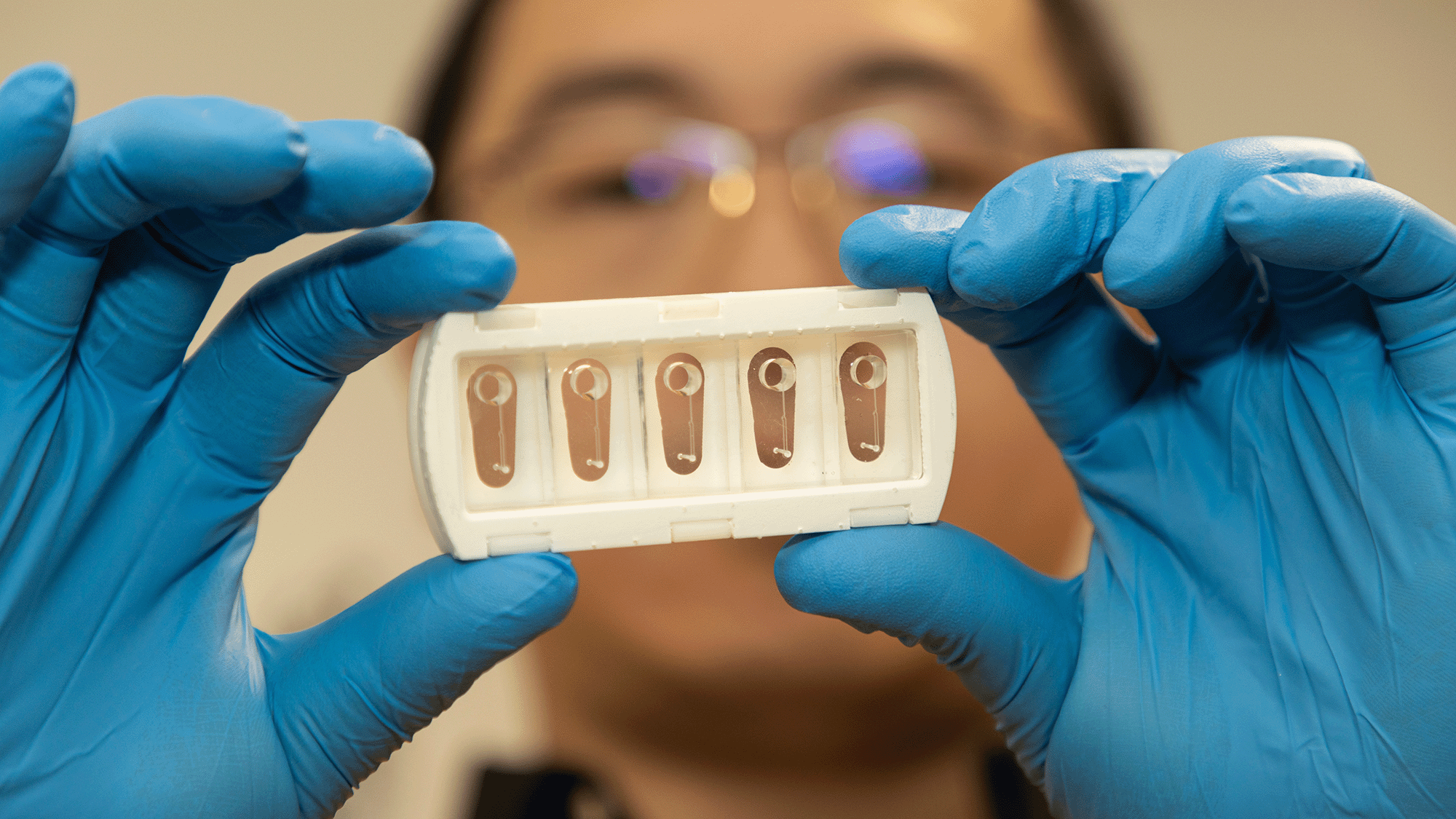
Recent advancements in vascular research have led to the development of miniature models replicating an individual's carotid arteries, offering valuable insights into stroke risk assessment. This research, featured in the journal Cell Biomaterials, demonstrates how these “arteries-on-a-chip” can mimic real blood flow, providing a unique platform for doctors to monitor clot behavior in patients.
Bridging Gaps in Patient Care
By utilizing this innovative approach, medical professionals may not only identify the nature and formation of clots in individual patients but also customize treatment plans based on real-time data. Charles Zhao, the study’s lead author and a PhD candidate at the University of Sydney, emphasizes the model’s relevance for complex cases where traditional imaging methods fall short, such as patients with recurrent strokes despite ongoing treatment. This situation is alarming but not uncommon in medical practice, where a one-size-fits-all approach often fails to account for individual variations in anatomy and physiology.
Precision medicine has been a key focus in recent years, and this new technique exemplifies how personalized diagnostics can potentially alter clinical outcomes. If you’re working in this space, the implications of these findings extend far beyond the research community; they suggest a future where treatment could be more effective and less reliant on generalized treatment protocols.
Understanding Ischemic Strokes
Ischemic strokes, responsible for a significant number of fatalities globally, typically arise from blood clots obstructing cerebral arteries. Current statistics show they account for about 87% of all strokes, underscoring their prevalence and the dire need for effective interventions. In Zhao's words, the presence of risk factors like obesity, hypertension, and high cholesterol highlights the need for advanced diagnostic techniques. Atherosclerosis, characterized by plaque accumulation, can lead to arterial damage, leaving tissues vulnerable to clot formation.
This dynamic creates a vicious cycle: as the clots form and propagate, the risk of subsequent strokes increases. The medical community has been aware of this link for years, but the challenge has consistently been accurately detecting and understanding these clots before they result in catastrophic outcomes. Current imaging methods are limited; they can pinpoint blocked arteries but often fail to reveal the nature and behavior of the clots—a critical gap in effective patient care.
The Mechanics of Clot Formation
When the inner lining of an artery sustains damage, essential materials beneath the cellular layer become exposed, triggering a reaction wherein a protein known as von Willebrand factor (VWF) interacts with platelets. This interaction initiates a series of aggregations, forming a clot. However, the real risk lies in the fate of this clot—whether it remains stationary or fragments and travels to the brain. The variabilities in an individual’s vascular makeup can influence this process, which traditional imaging cannot address.
Zhao points out that while current imaging methods excel at identifying arterial narrowing, they do not provide conclusive insights into clot behavior. This gap is where the “artery-on-a-chip” models are effective. These structures are created using patient-specific CT scans, which are 3D printed to mirror the precise anatomy of their carotid arteries. This specificity is significant because it allows for more accurate simulation of blood flow and clot behavior. Following that, a human collagen layer is applied to the model, and endothelial cells are carefully layered to simulate the inner artery lining. This meticulous approach ensures that the findings are not merely theoretical but can potentially guide real-world clinical practices.
Simulating Clot Development
Applying a laser injury allows researchers to expose the collagen and simulate thrombosis—blood clot formation—under controlled conditions, revealing critical insights into how clots develop within a given vascular context. This method is not just a technical feat; it’s a meaningful step towards understanding the complexities of clot behavior. The team analyzed specimens from six patients with varied arterial conditions, illustrating the significant differences in clot behavior due to unique blood flow patterns shaped by individual vessel anatomy.
This localized assessment is especially pertinent when strategizing stroke risk reduction. Various therapeutic approaches targeting platelets can be employed—some prevent aggregation, while others stop adhesion to arterial walls. The treatment landscape for stroke is crowded, but Zhao notes that the effectiveness of these treatments hinges on understanding the specific clot formation dynamics present in each patient. Tailoring interventions based on real-time data could fundamentally change how doctors approach stroke prevention.
Future Implications and Conclusions
Going forward, the research team aims to establish these artery-on-a-chip models as a standard diagnostic step that enhances decision-making between initial imaging assessments and treatment options. By focusing on patient-specific biological reactions within these models, clinicians may derive more tailored and effective interventions for stroke prevention in the future. The implications here are significant, as they could streamline treatment protocols dramatically.
This investigation offers a promising avenue for the advancement of personalized medicine, creating a framework that could eventually lead to improved outcomes for patients at risk of ischemic stroke. Such a leap forward would not just be a technical success—it could shift the way we think about patient care in vascular medicine.
Test your knowledge about cardiovascular health with our heart quiz!
Discussion
Sign in to join the discussion.